Fluorescein angiography
What is fluorescein angiography?
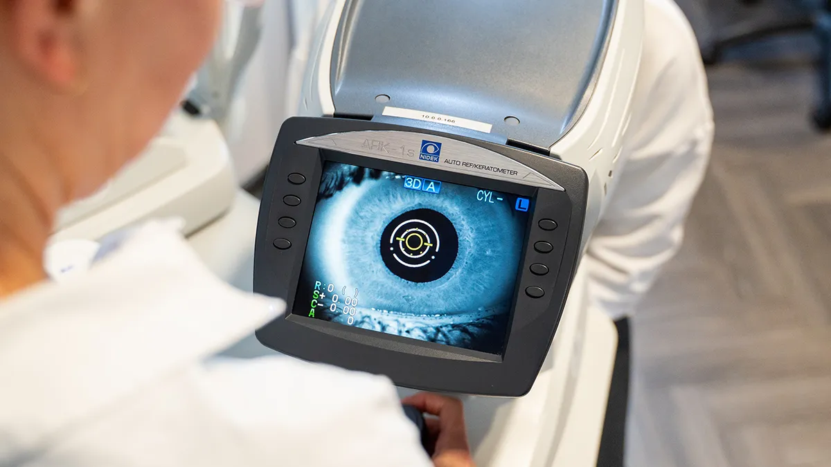
Fluorescein angiography is an imaging procedure for examining the fundus of the eye. A dye is administered intravenously, which allows us to take a closer look at the blood vessels at the back of the eye after a few seconds. This enables us to recognise pathological changes in the eye at an early stage. Before the treatment, special eye drops are administered to dilate the pupil so that the outer areas of the retina can also be viewed better. If it is not possible to dilate the pupil, we can also take an image of the outer areas of the retina using a wide-angle camera, which is available to you at BeyondEye. A special camera is then used to take a series of photos, which make it possible to analyse the blood flow in the eyes and the condition of the eye. The examination usually takes no longer than five minutes, as the dye in the eye is already broken down again after this time.
What are the risks and side effects of fluorescein angiography?
In rare cases, the mydriatic administered or the intravenously administered dye may cause intolerance reactions in the body. These may be characterised by nausea, dizziness or skin reactions. An allergic reaction can also lead to circulatory shock. Furthermore, the eye drops administered may cause an attack of glaucoma. A yellow colouration of the skin and urine regularly occurs after the examination and is no cause for concern. Serious complications are extremely rare in the vascular imaging procedure (angiography). Fluorescein angiography is a routine examination procedure in ophthalmology in Germany.
Important to know
Due to the pupil-dilating drops, patients have limited vision for several hours after the examination. They are therefore not allowed to actively participate in road traffic. Operating heavy machinery and carrying out dangerous activities are also prohibited. It is best if someone picks you up at the practice after the examination and takes you home.