Wide-angle photography and fundus photography
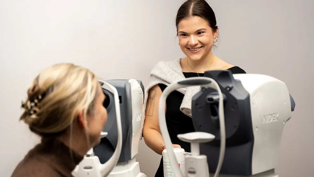
It is important to us at BeyondEye to always keep our practice at the cutting edge of technology. That's why we have the “California” from Optos. This enables us to take photographs, fundus autofluorescence images and fluorescein angiography images of the fundus of the eye. The special thing about it is that even without administering pupil-dilating eye drops, wide-angle photography - i.e. wide-angle images of the back of the eye up to the periphery - can be taken.
When are wide-angle photography and fundus photography useful?
These examinations can be used particularly well to document retinal degeneration, retinal detachment, choroidal birthmarks or diabetic retinopathy. Wide-angle fundus autofluorescence, for example, is ideal for examining hereditary retinal diseases such as retinitis pigmentosa. We use wide-angle fluorescein angiography for advanced diabetic changes in the fundus of the eye and retinal vascular occlusions. This enables us to diagnose circulatory disorders that affect the outer parts of the retina particularly well.